Structural heart disease (SHD)
A unified solution delivers valuable benefits to your team
A range of key capabilities across the SHD pathway
Elevate patient and staff experiences to provide better personalized care and enhanced outcomes
Cardiology departments need to be future-ready to face profound challenges
Demonstrated results in SHD care
Demonstrated results in SHD care
82% time saved for quantification of the LV and LA
Time acquisition and analysis for LV and LA alone was significantly faster than that of the 2DE images with the use of dynamic HeartModelA.I. 3D software for robust, reproducible ejection fraction (EF) as part of a routine workflow.1
52% reduction in radiation dose in LAA occlusion
This claim is based on a single center study. The 52% reduction is a comparison of radiation dose between two groups of patients (with (n=17) and without (n=17) echonavigator guidance). This claim is limited to left atrial appendage occlusion.2
99% success rate for Auto View Recognition
Auto View Recognition automatically identifies which selected image is apical 4 chamber (A4C), apical 2 chamber (A2C) and apical 3 chamber (A3C), and automatically assigns the labels to the selected images. The label is shown on the image as a schematic overlay. The algorithm has been validated on more than 6,000 clinical images with a success rate of 99%. This means that only one out of 100 cases will require manual intervention.3
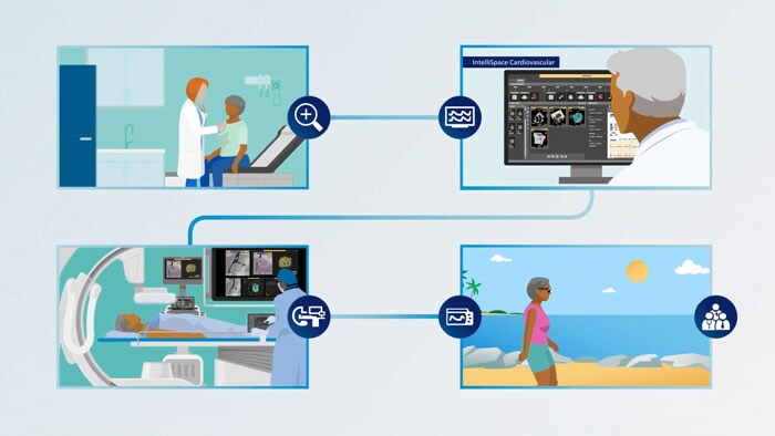
Care throughout the patient journey
Our solutions seamlessly connect to provide the right data at the right time—so you can diagnose with confidence, select the right treatment for each patient and move them efficiently through an interventional procedure while working to help support exceptional care.
Products and solutions for SHD
SHD Suite
Integrated solutions that seamlessly connect to bring your care to the next level.
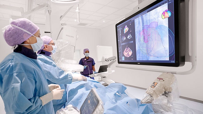
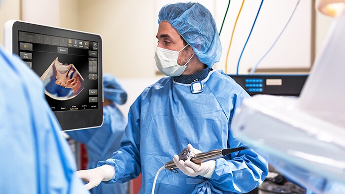
EPIQ CVxi ultrasound
EPIQ CVxi image quality and photorealistic imaging provide enhanced visualization along with automated quantification capabilities to help optimize device placement.
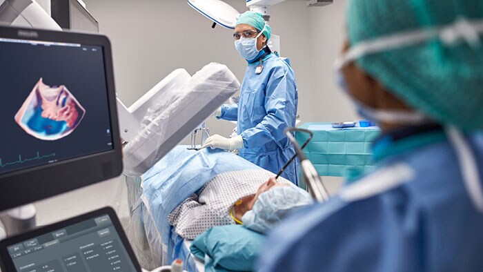
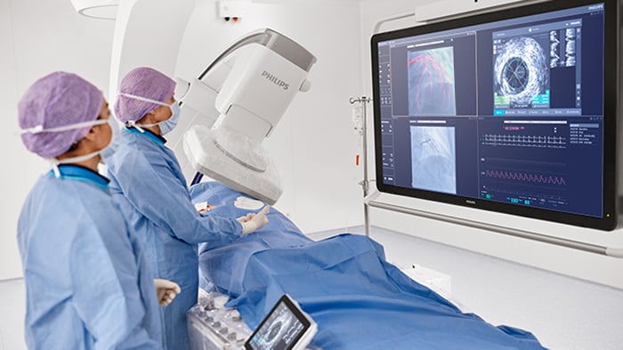
EchoNavigator
Our advanced EchoNavigator solution assists heart teams with fast, intuitive live fusion of X-ray and ultrasound imaging.

Cardiac imaging
Explore how imaging technology can help deliver life-changing clinical insights to patients and cardiology teams by providing exceptional quality in diagnostic imaging and streamlined analysis for a more confident diagnosis.
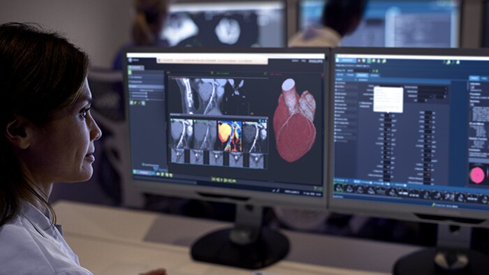
Advanced Visualization 15
Intelligent, automated and connected advanced visualization solution for all your analysis needs in one comprehensive solution.

"With the Philips lab, these procedures are so comfortable, so efficient - and the imaging is beautiful"
Lucy M. Safi, DO, FACC, FASE Director of Interventional Echocardiography
Hackensack University Medical Center
Hackensack, NJ, US
1/3
Customer story
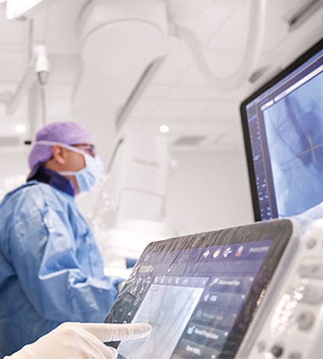
Establishing a Center of Excellence for structural heart disease
Establishing a Center of Excellence for structural heart disease
Discover how Hackensack University Medical Center has expanded its reputation as an academic medical center into a Center of Excellence for treating structural heart disease.
2/3
Customer story
Clinical confidence and efficiency in structural heart disease: an administrator’s perspective
Clinical confidence and efficiency in structural heart disease:
Hackensack’s Director of Network Operations for Cardiovascular Care, Hilary Nierenberg, FACHE, shares her perspective on Hackensack’s growth and the ways that Philips has helped support that growth. Read on to discover more.
3/3
Customer story
Clinical confidence and efficiency in structural heart disease: a physician's perspective
Clinical confidence and efficiency in structural heart disease:
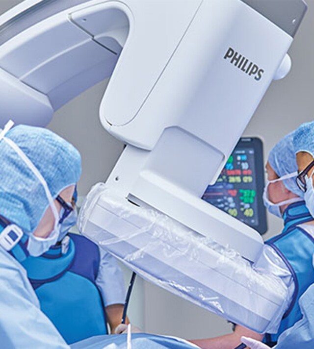
Discover what Hackensack’s lead interventional cardiologist and echocardiographer have to share about their experience with Philips integrated solutions in the lab and their outlook on what is to come in the future of SHD treatment.
References
2Jungen et al., PLoS One 2015 Oct 14;10(10); Left Atrial Appendage Closure Guided by Integrated Echocardiography and Fluoroscopy Imaging Reduces Radiation Exposure 3AutoStrain LV/RV/LA – automated strain measurements. Verena Roediger, PhD, Product Manager, TOMTEC.
